Ankle sprains are the most common issues in ankle injuries, typically occurring due to excessive inversion, causing the ankle to twist inward. Such sprains often result in injury to the ligaments that support the ankle from the outside.
Injury Severity
- Grade 1: Localized, mild swelling, and slight pain are present. The Anterior Talofibular Ligament (ATFL) is affected, with some tension in the ligaments. No sign of joint instability.
- Grade 2: Localized, moderate swelling and pain are present. Partial tears in the ligaments are observed. Walking normally or without crutches is difficult. Some mild joint movement abnormalities (instability) may be noticed.
- Grade 3: Widespread and significant swelling is present. Severe pain, tenderness, and loss of function are observed. The ligaments are completely torn. Joint movement abnormalities (instability) are evident.
Achilles Tendon Injury
This is a common sports injury in young and middle-aged athletes. However, it can also occur during simple household tasks, while going up and down stairs, or after a jump.
Causes of Achilles Tendon Injury
It can occur when the knee is extended, and trauma comes from above, or due to repeated microtraumas.
Symptoms
Local tenderness, a gap in the Achilles tendon, and ankle movement abnormalities may develop.
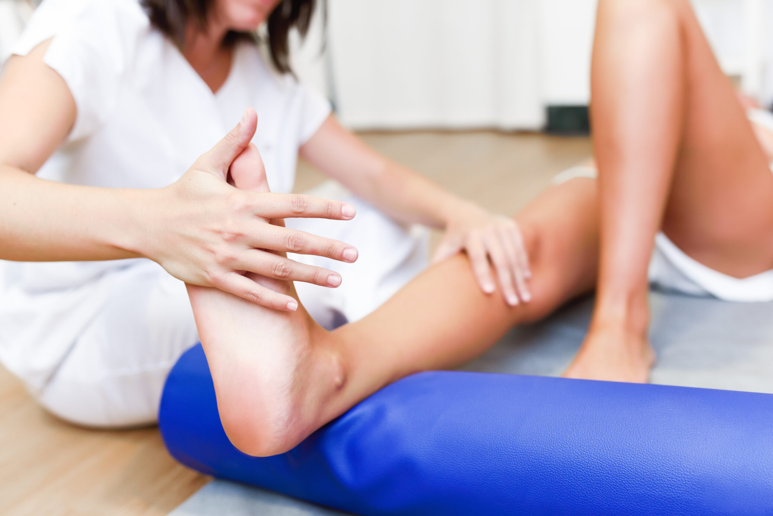
Treatment for Foot and Ankle Injuries
The initial weeks following an injury, trauma, or surgery constitute the early rehabilitation phase. In this stage, principles of protection, cold therapy, compression, and elevation should be applied.
Now, I’ll translate the text into German:
Fuß- und Knöchelverletzungen und ihre Behandlung
Probleme im Zusammenhang mit dem Fuß und dem Knöchel können bei Menschen jeden Alters und bei Sportlern aufgrund angeborener Faktoren, Traumata und Überanstrengung auftreten. Der Knöchel ist aufgrund der Anordnung seiner Knochen und Bänder strukturell stark und wird in Fällen von Überlastung in der Regel als erste Region verletzt, da er das gesamte Körpergewicht trägt.
Anatomie des Knöchels
Das Knöchelgelenk setzt sich aus mehreren Knochen, Bändern, Muskeln und Sehnen zusammen.
Das Knöchelgelenk besteht aus vier Knochen:
- Tibia (Schienbein)
- Fibula (Wadenbein)
- Talus (Sprungbein)
- Calcaneus (Fersenbein)
An beiden Seiten des Knöchels befinden sich gerundete Knochenstrukturen, die die entfernten Enden der Tibia (auf der Innenseite) und der Fibula (auf der Außenseite) sind. Diese Strukturen, als Malleoli bekannt, bilden auch die Anheftungspunkte für Muskeln, Sehnen und Bänder.
Die Bänder des Knöchelgelenks umfassen:
- Vorderes Talofibularband (ATFL)
- Calcaneofibulares Band
- Hinteres Talofibularband
- Deltaband
Diese Bänder sind besonders anfällig für Verletzungen bei Knöchelverstauchungen.
Frührehabilitation
Das Hauptziel in der Frührehabilitation besteht darin, das verletzte Gewebe vor weiteren Schäden zu schützen. Dies wird durch Methoden wie Tapeverband, Bandagen und Wickeln zum Schutz erreicht.
Der erste Schritt besteht darin, Schmerzen zu lindern und Schwellungen zu reduzieren. Lokale Kältetherapien und elektrische Ströme werden zur Schmerzlinderung verwendet. Um Schwellungen zu stoppen und zu verringern, werden Techniken wie JOBS (Intermittierende Kompressionsgeräte), Lymphdrainage, Matrixtherapie und Ultraschalltherap